animal cell microscope experiment
Comparing Plant and Animal Cells looks at cheek and onion. Microscope is used extensively in cell biology microbiology biotechnology microelectronics nanophysics pharmacology mineralogy and forensics.

Cells Microscope Activity Unit Animal Cell Cell Model Cells Project
Add a drop of purple stain specific for animals and cover with a cover slip.

. Students make a wet-mount slide of their own cheek cells and another of onion cells. Hold the coverslip or another slide with one end flush on the slide and gently wipe the edge of the coverslip over the scrapings. It is placed on the stage of the microscope.
Using the microscope features you will explore each slide specimen and. See also how to pass nclex rn after failing. Observe the cheek cells under both low and high power of your microscope.
This is called a wet mount. Set up your microscope. Part 1 - Animal Cells If we did this in class we would get cheek cells by scraping the inside of the mouth with a toothpick and then rubbing the toothpick on a drop of water with blue stain.
Cover slip A thin and transparent plastic slip to cover the specimen on the glass slide Safranin Used to stain the cells to be observed better under the microscope. Cells are taken from the inside cheek of a student. Scrape the inside of your cheek with the Q-tip and wipe it on to the center of the slide.
Examination of plant and animal cells under the microscope. We use microscope comprehensively in microbiology mineralogy cell biology biotechnology nano physics microelectronics pharmacology and forensics. Investigating cells with a light microscope.
List of Microscope Experiments for Kids 1. The virtual lab begins at the step where you place the slide on the microscope page. After hundreds of years of observations by.
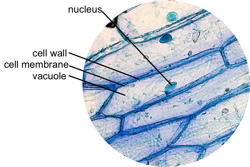
To investigate plant and animal cells using light microscopy. Aims of the experiment. Onion - A simple layer of onion skin is a great introduction to looking at plant cells.
Rinse off the stain and allow the slide to dry. Spider Web - Clear nail polish is all you need to see how amazing a spider web really is. Cheek Cell Lab observe cheek cells under the microscope Cheek Cell Virtual Lab virtual microscope view of cells.
Comparing Plant and Animal Cells. Gently tap the slide with a pencil to remove any air bubbles. For the Lesson 9 science experiment from the Botany unit from The Good an.
Place a drop of water on the slide. Plant Cell Lab microscope observation of onion and elodea Plant Cell Lab Makeup can be done at home or at the library Plant Cell Virtual Lab use a virtual microscope to view plant cells. This lab activity on viewing plant and animal cells under the microscope will prepare your 7th grade science and biology students to walk through the steps to set up and view samples of plant and animal cells properly.
Most of the structures within the cells using your microscope however you will be able to distinguish differences between the plant and animal cell and view a few of the larger organelles. In this investigation you will use a virtual microscope to view slides of cork cells onion bulb epidermis cells privet leaf cells and cheek cells. This discovery proposed as the cell doctrine by Schleiden and.
Place the onion skin in the center of the slide. In this simple microscope experiment we will compare plant cells and animal cells. To use a light microscope to examine animal or plant cells.
Observing a wide range of biological processes and animal cell under light microscope is easier due to advances in microscopic techniques. Distilled water To wet the specimen on the glass slide Razor knife Sharp knife to cut the onion. Animal and Plant Cells Microscope Lab Onion and Cheek Cells by Miss Middle School Teacher This is a quick and easy way to have students observe actual animal and plant cells.
Pond Water - Your favorite pond may be teeming with more life than you think. Ever since the first microscope was used biologists have been interested in studying the cellular organization of all living things. Place a small drop of methylene blue on a clean slide.
The students noticed the differences and similarities between the animal and plant cell under microscope. Put a couple of drops of methylene blue stain on the smear and leave for a few mintues. This practical involves looking at animal human cells through a microscope.
Students will compare the structures in. Using one of the cut sections of onions at your station remove the single layer of epidermal cells from the onion the thinner the better Place the layer of tissue on a slide and then add a. To prepare animal cells for viewing under a microscope.
Cheek Swab - Take a painless cheek scraping to view the cells in your own body. However plant and animal cells do not exactly look the same or do not. After the swabs and slides have been used they need to be soaked in disinfectant and disposed of according to CLEAPSS recommendations.
Structurally plant and anmimal cells are very similar because they are both eukaryotic cells. Place the two drops of water on the onion skin. Equipment required per set.
Get a clean slide. Students should only extract and view their own cells. Plant cells are eukaryotic cells where.
The plant cells contain chloroplast since they undergo photosynthesis but animal cells do not have this. Sterile cotton swabs Swabbed inside the cheeks to obtain cheek cells 70 RESULTS. Detailed aspects of the cell will be studied in the next section.
Use a cotton swab to get cheek cells and smear this onto a slide. It was not until good light microscopes became available in the early part of the nineteenth century that all plant and animal tissues were discovered to be aggregates of individual cells. A typical animal cell is 1020 μm in diameter which is about one-fifth the size of the smallest particle visible to the naked eye.
Once slides have been prepared they can be examined under a microscope. Starting at one edge gently lower a coverslip over the onion skin. This is called a smear and it makes a specimen layer thin enough to view clearly.
The blue helps you see the cells which are normally a clear color. Ive had quite a. While observing with tissues or on tissue.
ANIMAL CELLS INTRODUCTION Background Information. Gently roll and rub the toothpick onto the top of a glass slide in an area that will be visible through the microscope. A plant cell is the structural and functional basic unit of life in kingdom plantae.
It is also used for medical diagnosis particularly while dealing with tissues or in smear tests on free cells or tissue fragments.

Onion And Cheek Cell Lab Experiment Organelles Science Cells Biology Classroom Life Science Middle School

Epidermal Onion Cells Under A Microscope Plant Cells Appear Polygonal From The Cell Diagram Plant Cell Diagram Plant Cell

Cells Microscope Things Under A Microscope Cell Plant And Animal Cells

Cell Lesson Cheek And Onion Cell Cell Theory Cells Lesson Cell Theory Biology Units

Structure Of Animal Cell And Plant Cell Under Microscope Diagrams Cell Diagram Plant Cell Diagram Animal Cell

Cells Observation Lab Activity Plant Cell Lab Activities Cells Worksheet

Biology Lab Variation In Cell Structure Plant And Animal Cells Biology Labs Biology

Microscope Lab Templates Plant And Animal Cells Science Projects For Kids Middle School Science Resources

Onion Cells Under A Microscope Requirements Preparation Observation Plant And Animal Cells Animal Cell Plant Cell